
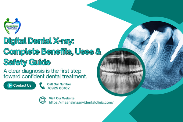
Digital Dental X-ray is a state of the art digital imaging system to assist your dentist in viewing areas of your mouth that are usually not accessible through a simple clinical examination. It may assist with the diagnosis and treatment of cavities, infections, bone changes, impacted teeth, and other dental issues.
We live in an era of modern dentistry, where dental X-rays are a crucial component as some dental problems may occur under the surface of teeth or within jawbone. Imaging adds pertinent data, which complemented with a clinical exam narrows the understanding of a patient’s oral health.
What is Digital X-ray in Dentistry?
A Digital Dental X-ray is a type of x-ray which utilizes electronic sensors to record images of teeth, root and surrounding bone along with other oral structures. A digital screen or computer then displays the image that allows the dental team to focus in a particular area for better evaluation.
Digital imaging does not require film processing with chemical-based X-rays. Being digital images, these can also be saved electronically giving them the added benefit of being easily maintained for keeping track of dental records and examining past records if need be.
The type of X-ray deemed necessary depends on the patient’s symptoms, dental history, age and oral health as well as the treatment being considered.
Why Are Dental X-rays Needed?
A dental examination involves more than simply looking at the visible surfaces of the teeth. Some problems can develop between teeth, around tooth roots, beneath existing restorations, or inside the jawbone.
A Digital Dental X-ray may help a dentist evaluate:
- Tooth decay between teeth or below existing fillings
- The condition of tooth roots
- Bone levels around teeth
- Impacted or developing teeth
- Dental infections and changes around tooth roots
- Certain tooth fractures
- Jawbone structures
- Teeth before extraction
- Teeth requiring root canal treatment
- Areas being evaluated for orthodontic or implant treatment
The dentist decides whether an X-ray is necessary based on the patient’s individual condition rather than simply taking an image routinely.
Types of Digital Dental X-rays
Types of Dental X-Rays Each type provides a different view of the mouth with some more focused than others.
Intraoral X-rays
They are taken within the mouth and can provide close-up views of single teeth, roots, and supportive bone. Some common types of X-rays which are used for specific diagnosis include bitewing and periapical X-ray.
Panoramic X-rays
Panoramic pictures are wider views of the teeth, jaws and nearby structures. Useful in assessment of wisdom teeth, impacted teeth and jaw anatomy or in treatment planning for some procedures.
The imaging technique is based on what has to be examined by the dentist. However, this does not hold true for every patient needing all types of dental X-rays.
How Does Digital X-ray Work at a Dental Office?
The process is generally quick. The dentist places a digital sensor either inside or outside the mouth, depending on what type of image is needed. This means you may need to hold the position or be required to bite on a positioning device while your image is taken.
This information is registered by the sensor, and that will be transmitted to a computer system or digital imaging system. The dentist will then be able to review this image in combination with the clinical examination.
A Digital Dental X-ray is especially useful because digital images can be blown up right on a screen for the dental staff to closely examine details in certain areas.
Benefits of Digital Dental X-ray
In modern dentistry, with digital dental imaging there are many practical advantages.

Quick Image Availability
In the case of digital images, these are universally to be looked at immediately after shooting. This can facilitate an effective examination and conversation between the dentist and patient.
Additional Diagnostic Information
X-rays are able to show structures that are not actually visible with a full view only. They may assist in the discovery of undiscernible oral issues and also aid in therapy preparation.
Easy Digital Storage
It is also possible for digital images to be stored electronically as part of a patient’s dental records, following effective privacy and record-management procedures.
Useful for Treatment Planning
These X-rays can help you plan on procedures such as root canal treatment, tooth extraction, orthodontic treatment and dental implant assessment.
Convenient Image Review
Invisible problems the kind that you may never find on a regular radiograph—can be visualized easier, digital images can be shown on the computer screen and blown up when necessary to assist the dental team in their investigations of specific areas.
Is Digital Dental X-ray Safe?
Dental X-ray: Dental X-rays are ionizing radiation, should be taken only when clinically warranted. Modern imaging procedures are used to get clinically relevant diagnostic data while minimizing X-ray exposure.
The dentist then recommends an X-ray after factors such as symptoms, age, dental history, and previous images which would guide the treatment decisions.
Tell your dentist if you are pregnant or could become pregnant, PRIOR to X-ray. That thereby enables the dental team to take a proper decision on whether imaging is needed.
When Might You Want to Use a Digital Dental X-Ray?
In some cases, your dentist may suggest imaging if you have symptoms or clinical findings that warrant more information.
Digital Dental X-ray may help with:
- Investigating unexplained tooth pain
- Checking suspected cavities
- Evaluating a dental infection
- Assessing impacted wisdom teeth
- Planning root canal treatment
- Planning tooth extraction
- Evaluating teeth before orthodontic treatment
- Assessing supporting bone
- Monitoring certain dental conditions
- Planning selected restorative procedures
An X-ray is a diagnostic aid rather than a treatment by itself. The dentist interprets the image together with your symptoms, medical and dental history, and clinical examination.
Digital Dental X-ray at Maansi Maanvi Multispeciality Dental Clinic
Maansi Maanvi Multispeciality Dental Clinic goes on to explain general dental procedures on its website, which includes dental exams and X-rays. The website also lists digital dental diagnosis as part of its approach for dental care.
Digital Dental X-ray may be done at Maansi Maanvi Multispeciality Dental Clinic if the dentist needs to have more information in order to diagnose a condition or plan treatment. Whether it is panoramic or 3D imaging, the imaging technique should be tailored to each individual patient.
General dentistry, oral surgery, endodontics, orthodontic treatment, dental implants and crowns and bridges are among the services offered by the clinic. Many of the imaging modalities are relevant to diagnosis or treatment planning of dental conditions.
Getting Ready for a Dental X-ray
Before dental imaging, little or no preparation is usually needed. You should wear comfortable clothes and listen to directions given by the dental team.
Inform your dentist about:
- Pregnancy or possible pregnancy
- Previous dental X-rays
- Existing dental conditions
- Relevant medical history
- Any removable dental appliances
You will also be asked to take off jewellery, removable dental appliances or similar objects that could affect image quality.
What Happens After the X-ray?
Once the images are taken, your dentist will review them with your clinical examination.
If the image reveals a potential issue, your dentist can explain what it might indicate and options for effective treatment. In some cases an X-ray also does not display a significant abnormality which itself provides useful information in the exploration of dental symptoms.
The decision for the next step will depend on the total clinical picture.
Frequently Asked Questions
What about Digital Dental X-ray Pain?
No. Dental x-rays do not involve cutting or drilling. A few intraoral sensors may even be positioned for a bit of time so it can feel slightly uncomfortable.
How frequently should dental X-rays be taken?
There is no one-time-table-fits-all. The frequency is based on your age, oral health, signs and symptoms, the history of the dental home, how risky the person will be in getting disease and clinical judgment by the dentist.
Can X-rays detect cavities?
Yes. Dental X-rays can detect cavities in parts of the mouth that are hard to assess with a normal visual inspection, especially between teeth.
Do children need dental X-rays?
If a child requires X-rays for clinical purposes, particularly when monitoring developing teeth, detecting cavities, or assessing orthodontic issues. A dentist will decide if imaging is needed.
Can a dental examination be replaced by an X-ray?
So, if you are familiar with X-rays. No. When required, a thorough evaluation may comprise medical history, dental | clinical examination and other tests.
Conclusion
Digital Dental X rays offer different insights into teeth, roots, bone and more that may be not as straightforward to see during a normal exam. Where appropriate, it enables dentists to diagnose or plan for possible treatment.
Maansi Maanvi Multispeciality Dental Clinic lists dental exams & X-Rays under General dentistry general procedures on its website, while digital dental diagnosis is also mentioned as part of an approach to patient care.
Is dental imaging right for your condition?If you have persistent tooth pain, swelling, sensitivity, a suspected cavity, impacted teeth or treatment ahead of you from the dentist such as surgery for wisdom teeth extraction or to help in preparation for securing implants and other delicate procedures that could also involve use of special equipment.